Regenerative Medicine for Canine Cartilage Disorders
Musculoskeletal disease is a common concern for working and performance dogs, as well as all aging dogs. The relatively new field of regenerative medicine offers exciting treatment options for these patients. By using the body’s own cells and proteins to promote healing, regenerative medicine may be used to treat tendon and ligament injuries, spinal conditions, and osteoarthritis in dogs. One of the ways AKC Canine Health Foundation (CHF) has invested in the study of regenerative medicine to treat canine osteochondrosis is through Grant 02078: Development of a Regenerative Medicine Technique to Treat Cartilage Disorders in Dogs. With this funding, researchers at Texas A&M University are discovering new ways to treat this painful cartilage disorder.
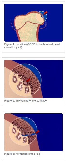
Osteochondrosis is an abnormal development of the cartilage lining the joint at the end of a bone (Figures 1-3). Immature cartilage fails to form bone and becomes thickened. The thickened, abnormal cartilage can separate from the underlying bone, creating a flap. Osteochondritis Dissecans (OCD) is the inflammatory condition that develops when this cartilage flap detaches, becoming a “joint mouse” and leaving an osteochondral defect that can progress to osteoarthritis. While regenerative medicine may offer a treatment alternative for this osteochondral defect, the unique interface of cartilage and bone has presented a challenge to the development of usable tissue scaffolds. The three tissue regions of this interface (cartilage, calcified cartilage, and subchondral bone), each have distinct extracellular matrix components and cell types that must be recreated for successful regenerative therapy.
CHF-funded researchers first set out to optimize the materials used to create an osteochondral tissue scaffold. By combining two tissue engineering techniques, they were able to produce a greater variety of tissue scaffold characteristics (such as pore size and pore interconnectivity) compared to conventionally available scaffolds. Since ideal scaffold characteristics are tissue-specific, having more tissue scaffold options may lead to more success in regeneration at the osteochondral interface.
Next, they explored the biochemical cues needed in a tissue scaffold to successfully direct synovium-derived stem cells (those harvested from a joint) to differentiate into the three layers of the bone-cartilage interface necessary for osteochondral repair. Researchers tested four different biochemical cues in various combinations and were able to create the desired cell differentiation into cartilage, calcified cartilage, and bone when cultured separately.
Results of this research have been published in Macromolecular Materials and Engineering and the Journal of Biomedical Materials Research Part B. The next steps in this research will assess how the tissue constructs behave when cultured together, with the eventual goal of clinical application in the treatment of osteochondrosis and osteoarthritis. CHF and its donors continue to support this ongoing research with an educational grant to Dr. Shelby Gasson through CHF’s Clinician-Scientist Fellowship Program. By funding cutting edge research and supporting the next generation of canine health researchers, CHF remains committed to better treatments, more accurate diagnoses, and an improved understanding of the mechanisms that cause disease in dogs. Learn more about our work for dogs at akcchf.org.